Análise editorial
Por que importa
A RNFL que caiu pode ser o filme, não o nervo.
O número da RNFL decide se o glaucoma “progrediu”. O mesmo paciente costuma chegar com colírio crônico e superfície irregular. Se o scan ruim afina o número, a visita seguinte parece piora.
Ressecamento degrada o sinal. No Stratus e no Cirrus, sinal baixo devolve RNFL mais fina. No Spectralis a direção inverte.
Stein é time-domain, olho tapado, sem DED.
Evidência
Evidência
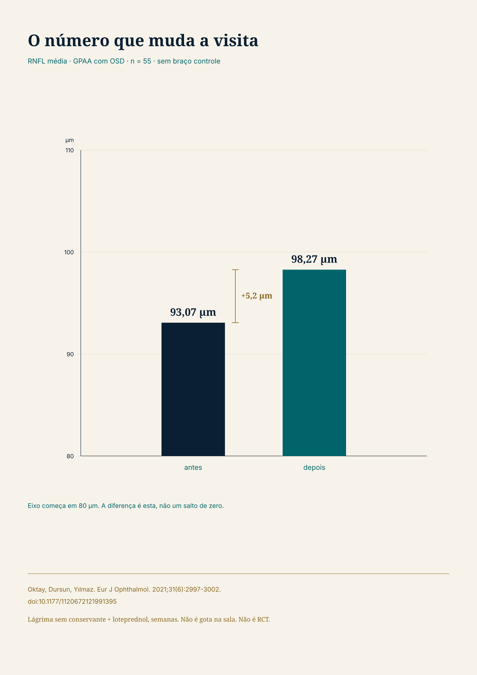
O número que muda a visita. RNFL média em GPAA com OSD, n = 55, sem braço controle: 93,07 µm antes → 98,27 µm depois de tratar a superfície (Δ +5,2 µm; eixo do gráfico 80–110 µm; lágrima + loteprednol, semanas; não é gota na sala; não é RCT). Oktay, Dursun, Yılmaz. Eur J Ophthalmol. 2021;31(6):2997-3002. doi:10.1177/1120672121991395
Stein e colaboradores (2006): 17 sadios, Stratus, pálpebra tapada. A RNFL cai. A queda passa o IC95% do erro depois de ~15 s. É time-domain, olho tapado, sem DED.
Cheung e colaboradores (2008) e Wu e colaboradores (2009): signal strength <7 afina a RNFL no Stratus. Huang e colaboradores (2011): Topcon e Cirrus.
Yohannan e colaboradores (2020): de signal strength 10 para 3, o Δ é pequeno. Abaixo de 3: −15,70 a −16,34 µm por ponto. Artefato: −4,76 µm.
Gershoni e colaboradores (2022) e Strampe e colaboradores (2020): no Spectralis, Q baixo engrossa a RNFL.
Asrani e colaboradores (2010): artefatos; olho seco tem mais artefato macular. Kesimal e colaboradores (2026): DED baixa o SSI no RTVue; o resumo não traz delta de RNFL em mícrons.
Oktay, Dursun e Yılmaz (2021): 55 GPAA com OSD; lágrima + loteprednol, semanas. Sinal 19,15→23,93. RNFL média 93,07→98,27 µm. doi:10.1177/1120672121991395. Não é gota na sala do aparelho. Não é RCT.
Hannemann e Erb (2021): gota de ácido hialurônico em sadio — OCT inalterado. Ghazi e Much (2009): n = 13 scans que falharam; o lubrificante subiu o sinal.
Não há claim de GCC. Não há claim de IA que calibra filme.
Aplicação
Prática

O que muda na cadeira. Piscar: quinze segundos de secagem já saem do erro do Stratus. Ler o sinal no eixo daquele aparelho — Stratus e Cirrus não são Spectralis. Tratar OSD e repetir: não chamar progressão no scan seco. Scan ruim não fecha piora, nem no sentido errado da plataforma.
Na cadeira, o número só entra na decisão depois do sinal e da superfície.
- Piscar: quinze segundos de secagem já saem do erro do Stratus.
- Ler o sinal no eixo daquele aparelho. Stratus e Cirrus não são Spectralis.
- Não chamar progressão em OSD sem tratar (Oktay ~5 µm).
- Gota imediata em sadio não puxa o número.
- Não misturar corte de sinal entre plataformas. IA não calibra filme nesta edição.
Limitações
Limitações
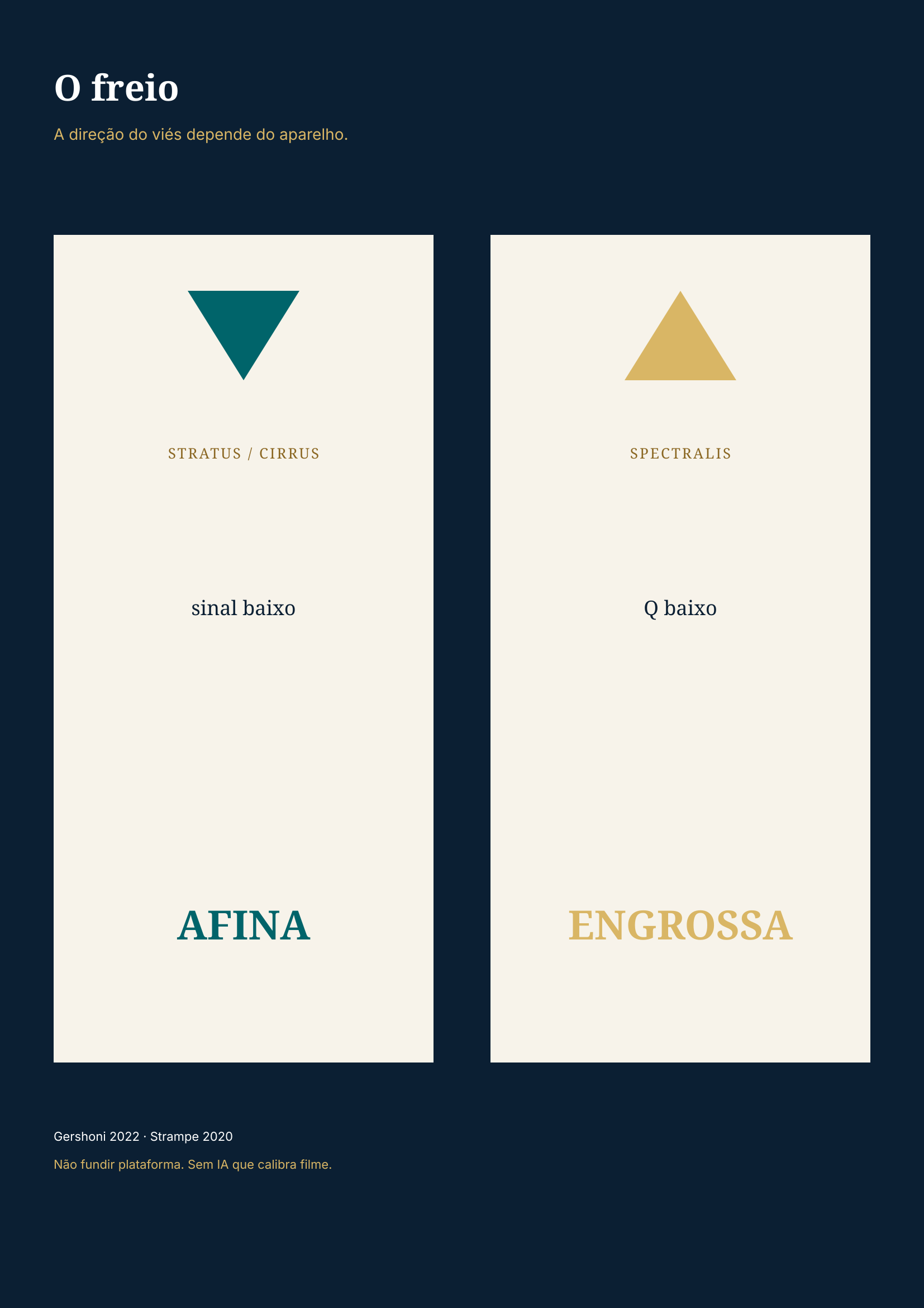
O freio: a direção do viés depende do aparelho. Stratus e Cirrus: sinal baixo afina RNFL. Spectralis: Q baixo engrossa. Gershoni 2022, Strampe 2020. Não fundir plataforma. Sem IA que calibra filme.
Stein é experimental.
Oktay: n = 55, um centro, sem braço controle.
O viés do Spectralis é o inverso do Cirrus e do Stratus.
Não há DOI de IA que calibra filme.
Fontes
Referências
- Stein DM, Wollstein G, Ishikawa H, Hertzmark E, Noecker RJ, Schuman JS. Effect of corneal drying on optical coherence tomography. Ophthalmology. 2006;113(6):985-991. DOI 10.1016/j.ophtha.2006.02.018 — Stein DM, Wollstein G, Ishikawa H, Hertzmark E, Noecker RJ, Schuman JS. Effect of corneal drying on optical coherence tomography. Ophthalmology. 2006;113(6):985-991.
- Cheung CYL, Leung CKS, Lin D, Pang CP, Lam DSC. Relationship between retinal nerve fiber layer measurement and signal strength in optical coherence tomography. Ophthalmology. 2008;115(8):1347-1351. DOI 10.1016/j.ophtha.2007.11.027 — Cheung CYL, Leung CKS, Lin D, Pang CP, Lam DSC. Relationship between retinal nerve fiber layer measurement and signal strength in optical coherence tomography. Ophthalmology. 2008;115(8):1347-1351.
- Wu Z, Huang J, Dustin L, Sadda SR. Signal strength is an important determinant of accuracy of nerve fiber layer thickness measurement by optical coherence tomography. J Glaucoma. 2009;18(3):213-216. DOI 10.1097/IJG.0b013e31817eee20 — Wu Z, Huang J, Dustin L, Sadda SR. Signal strength is an important determinant of accuracy of nerve fiber layer thickness measurement by optical coherence tomography. J Glaucoma. 2009;18(3):213-216.
- Huang J, Liu X, Wu Z, Sadda S. Image quality affects macular and retinal nerve fiber layer thickness measurements on Fourier-domain optical coherence tomography. Ophthalmic Surg Lasers Imaging. 2011;42(3):216-221. DOI 10.3928/15428877-20110324-01 — Huang J, Liu X, Wu Z, Sadda S. Image quality affects macular and retinal nerve fiber layer thickness measurements on Fourier-domain optical coherence tomography. Ophthalmic Surg Lasers Imaging. 2011;42(3):216-221.
- Yohannan J, Cheng M, Da J, et al. Evidence-based criteria for determining peripapillary OCT reliability. Ophthalmology. 2020;127(2):167-176. DOI 10.1016/j.ophtha.2019.08.027 — Yohannan J, Cheng M, Da J, et al. Evidence-based criteria for determining peripapillary OCT reliability. Ophthalmology. 2020;127(2):167-176.
- Gershoni A, Barayev E, Vainer I, et al. Thickness measurements taken with the spectralis OCT increase with decreasing signal strength. BMC Ophthalmol. 2022;22:148. DOI 10.1186/s12886-022-02356-4 — Gershoni A, Barayev E, Vainer I, et al. Thickness measurements taken with the spectralis OCT increase with decreasing signal strength. BMC Ophthalmol. 2022;22:148.
- Strampe MR, Eldweik L, Chaon BC, et al. Retinal nerve fibre layer thickness increases with decreasing Spectralis OCT signal strength in normal eyes. Neuroophthalmology. 2020;44(2):100-103. DOI 10.1080/01658107.2019.1653934 — Strampe MR, Eldweik L, Chaon BC, et al. Retinal nerve fibre layer thickness increases with decreasing Spectralis OCT signal strength in normal eyes. Neuroophthalmology. 2020;44(2):100-103.
- Asrani S, Edghill B, Gupta Y, Meerhoff G. Optical coherence tomography errors in glaucoma. J Glaucoma. 2010;19(4):237-242. DOI 10.1097/IJG.0b013e3181b21f99 — Asrani S, Edghill B, Gupta Y, Meerhoff G. Optical coherence tomography errors in glaucoma. J Glaucoma. 2010;19(4):237-242.
- Kesimal B, Kocamış Sİ. Investigation of the impact of tear film dynamics on optical coherence tomography image quality in patients with dry eye disease. BMC Ophthalmol. 2026;26. DOI 10.1186/s12886-026-04893-8 — Kesimal B, Kocamış Sİ. Investigation of the impact of tear film dynamics on optical coherence tomography image quality in patients with dry eye disease. BMC Ophthalmol. 2026;26.
- Oktay Ö, Dursun Ö, Yılmaz A. The effects of ocular surface disease on optical coherence tomography test results in patients with glaucoma. Eur J Ophthalmol. 2021;31(6):2997-3002. DOI 10.1177/1120672121991395 — Oktay Ö, Dursun Ö, Yılmaz A. The effects of ocular surface disease on optical coherence tomography test results in patients with glaucoma. Eur J Ophthalmol. 2021;31(6):2997-3002.
- Hannemann E, Erb C. The impact of artificial tear viscosity on the results of optical coherence tomography. Klin Monbl Augenheilkd. 2021;238(9):1004-1009. DOI 10.1055/a-1179-0131 — Hannemann E, Erb C. The impact of artificial tear viscosity on the results of optical coherence tomography. Klin Monbl Augenheilkd. 2021;238(9):1004-1009.
- Ghazi NG, Much JW. The effect of lubricating eye drops on optical coherence tomography imaging of the retina. Digit J Ophthalmol. 2009;15(2):24-26. DOI 10.5693/djo.01.2009.006 — Ghazi NG, Much JW. The effect of lubricating eye drops on optical coherence tomography imaging of the retina. Digit J Ophthalmol. 2009;15(2):24-26.
Transparência
Conflitos de interesse / disclosures
- Financiamento
- Sem financiamento externo.
- Conflitos de interesse
- O autor declara não possuir vínculo com fabricantes de dispositivos, fármacos ou tecnologias citados neste artigo.
- Uso de inteligência artificial
- Houve assistência de IA na reorganização editorial, na revisão linguística e na conferência bibliográfica das referências contra Crossref e PubMed. A IA não foi tratada como fonte; as afirmações e referências foram verificadas pelo autor.
- Revisão
- Este artigo não passou por revisão independente por pares. Ver o selo editorial no topo da página.
- Aprovação ética
- Não se aplica: revisão narrativa sem dados individuais ou imagens identificáveis.